The Microstructure of Poly(ethyl cyanoacrylate)
The Microstructure of Poly(ethyl cyanoacrylate)
Folder:
Journal:
Year:
DOI:
10.1002/pol.1988.140261102
Type of document:
Language:
The Microstructure of Poly(ethyl cyanoacrylate)
ALLAN H. FAWCETI', Department of Pure and Applied Chemistry, JOHN
GUTHRIE,* MIKE S. OTTERBURN: and DANNY Y. S. SZETO,
Department of Chemical Engineering, The Queens University of Belfast,
United Kingdom
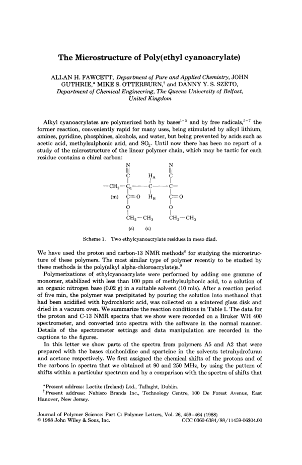
Alkyl cyanoacrylates are polymerized both by bases'-5 and by free r a d i ~ a l s , ~ - ~
the
former reaction, conveniently rapid for many uses, being stimulated by alkyl lithium,
amines, pyridine, phosphines, alcohols, and water, but being prevented by acids such as
acetic acid, methylsulphonic acid, and SO,. Until now there has been no report of a
study of the microstructure of the linear polymer chain, which may be tactic for each
residue contains a chiral carbon:
H,
C=O
I
CH2- CH,
(m)
C=O
I
CH,-
(4
CH,
(4
Scheme 1. Two ethylcyanoacrylate residues in meso diad.
We have used the proton and carbon-13 NMR methods8 for studying the microstructure of these polymers. The most similar type of polymer recently to be studied by
these methods is the poly(alky1 alpha-chloroacrylate)s?
Polymerizations of ethylcyanoacrylate were performed by adding one gramme of
monomer, stabilized with less than 100 ppm of methylsulphonic acid, to a solution of
an organic nitrogen base (0.02 g) in a suitable solvent (10 mls). After a reaction period
of five min, the polymer was precipitated by pouring the solution into methanol that
had been acidified with hydrochloric acid, was collected on a scintered glass disk and
dried in a vacuum oven. We summarize the reaction conditions in Table I. The data for,
the proton and C-13 NMR spectra that we show were recorded on a Bruker WH 400
spectrometer, and converted into spectra with the software in the normal manner.
Details of the spectrometer settings and data manipulation are recorded in the
captions to the figures.
In this letter we show parts of the spectra from polymers A5 and A that were
2
prepared with the bases cinchonidine and sparteine in the solvents tetrahydrofuran
and acetone respectively. We first assigned the chemical shifts of the protons and of
the carbons in spectra that we obtained at 90 and 250 MHz, by using the pattern of
shifts within a particular spectrum and by a comparison with the spectra of shifts that
*Present address: Loctite (Ireland) Ltd., Tallaght, Dublin.
'Present address: Nabisco Brands Inc., Technology Centre, 100 De Forest Avenue, East
Hanover, New Jersey.
Journal of Polymer Science: Part C: Polymer Letters, Vol. 26, 459-464 (1988)
0 1988 John Wiley & Sons, Inc.
CCC 0360-6384/88/11459-06$04.00
FAWCETT ET AL.
460
TABLE I
Reaction conditions for preparation of the polymer samples
~~~
~
Yield
Sample
Base
Solvent
g
M,t
4
'
A5
A2
Cinchonidine
Sparteine
tetrahydrofuran
acetone
0.92
0.90
229,000
153,000
0.54
0.68
'M, from styrene-calibrated GPC column, with THF as solvent.
*Calculated with the Bernoullian probability relationships of reference 8.
we have obtained of other alkyl cyanoacrylate polymer. We have entered the shifts in
Table 11. The chains consisted, as far as could be determined, of head to tail sequences
of the one residue. The signals from the main chain methylene and quaternary carbons
apparently overlap near 44 ppm, the former having a larger dispersion of shifts. The
fine structure of this region and of most of the other carbons w l be discussed in
i
l
another place; here we confine ourselves to the fine structure of the side chain
methylene carbon (Fig. 1) and the fine structure of the proton spectrum of the main
chain methylene protons (Fig. 2), which have been recorded at high field. It can be seen
that these parts of the spectra are sensitive to the method of preparation: in each
figure, parts (a) and (b) clearly differ in the relative intensities within a pattern of the
individual components. For the two preparations, either, and most simply the difference of solvent, or the difference of c h i d base is responsible for the difference in the
polymer microstructure that the spectroscopy has detected.
The 100 MHz spectra of the side chain methylene carbons from both poly(ethy1cyanoacrylate)s, as seen in Figure 1 parts (a) and (b), have three well-resolved peaks.
Some of these show shoulders or other features that became peaks upon resolution
enhancement (parts c and d) by modifying the FID prior to the Fourier transform, in
the manner that is indicated in the caption to the figure. As is appropriate and most
simple for a carbon that is attached to one main chain chiral atom and is located
midway between two others (7), we regard the three peak pattern as deriving from
stereochemical triads, the approximate 1 : 2 : 1 ratio of polymer A5 indicating approximate atacticity and the very different pattern of polymer A2 indicating a bias
towards tacticity of some kind, for the down-field peak is much reduced in intensity,
and the upfield peak is of slightly greater magnitude than the central heteroatactic
peak. The classical NMR approach to tacticity assignment is based upon the proton
spectrum of the main chain methylene groups, to which we now turn (Fig. 2).
If the m2thylene group proton signal showed diad sensitivity: we would expect a
single peak from the racemic or syndiotactic dyad and an AB or AX quartet from the
meso or isotactic dyads (with a coupling constant of about - 14 Hz," a spacing which
is indicated by a bar on the figure). Since there are more than 5 peaks or shoulders, the
'H
UC
166.2
115.7
2.6-2.St
44
44
4.26
64.41
1.32
13.7
*Shifts were measured in ppm relative to TMS at 12OOC for protons in dimethyl sulfoxide-d,
and 26°C for carbons in acetone-d,. The atoms are labeled in Scheme 1.
'The fine structure of these nuclei is shown in the figures and is discussed in this paper.
POLY(ETHYL CYANOACRYLATE)
461
rm
I
1
65
64
6 )P P ~
Fig. 1. Carbon-13 NMR spectrum of the side chain methylene groups of poly(ethy1
cyanoacrylate) a t 100 MHz. Sample A5 was used for parts (a) and (c), and sample A2 was used for
parts (b) and (d). The samples (about 200 mg) were dissolved in acetone-d6 at 299°K: PW 12 p s ,
AQ 0.66 ms, NS 2048. For parts (a) and (b) the FID was multiplied by an exponential function
with an LB of - 3.0 Hz,and a Gaussian Broadening factor of GB = 0.2 was used: for parts (c) and
(d), LB was - 5.0 Hz and GB was 0.35, to enhance the resolution further.l0
system displays at least partial tetrad sensitivity. This we now attempt to analyze for
the purpose of identifying the type of tacticity that predominates.
We have labeled the peaks at 2.75 and 2.70 ppm a and b respectively, and have
distinguished them because they are apparently single line features: their width at half
height is 9 or 10 Hz,they are separated by 20 Hz from each other and by at least 18 Hz
from the nearest other feature. They are not readily interpreted in terms of parts of
one or more AB systems (except in the sense that AuAB is vanishingly small), and so
probably derive from r-centred diads. The smaller peak, a, may be from the rrr
sequence, and the larger peak, b, may be from rrm,mrr sequences, whose probability
will be larger than the rrr sequence, if, as seems appropriate, the chains are mainly
isotactic. T h e features to the left and right of the pattern are clearly composed of a
number of multiplet l n s as is appropriate for a meso dyad assignment. It is the small
ie,
values of the relative areas of the a and b peaks within the patterns which indicate
that both polymers may be predominantly isotactic. The labels rr, rm/mr, and mm
have thus been attached to the I3C spectrum of Figure l(a) and (b), the smaller, low
field, signal being judged to be from the rr triad and the larger upfield signal from the
mm triad. From the relative areas of these, by assuming Bernoullian statistics: values
462
FAWCETT ET AL.
64.63
C)
d)
mm
rm
mr
I
64.90
I
I
I
6 P
P
1
64
1
65
65
I
I
I
64
~
Fig. 1. (Continuedfrom thepreoiouspage.)
of a single probability parameter, for isotactic placement,
p
,
=
(1
-
c,have been obtained. Thus
e)2, and so on8
We now re-examine the spectra to see how well the h e structure is consistent with
this interpretation. It may be seen, from the entries in Table I11 that the triad
probabilities, obtained for the triad features of the side chain methylene group’s C-13
spectra of Figure 1, are predicted well by this model for both polymers: there is no
strong indication of the need of a Markov model, with a second parameter, being
required. The tetrad assignments that we have made of the main chain methylene
protons are in good agreement, and the three rr-centred pentads of the side chain
methylene are quite well reproduced.
These two polymers show a slight and a moderate bias towards isotacticity. While it
is possible that a chiral anion, derived bom the chiral base and associated by
coulombic effects with the propagating
might promote the formation of an
isotactic, rather than a syndiotactic polymer, it may be that the simplest explanation
of the bias towards isotacticity is appropriate: that it is controlled by the process of
adding the monomer to the unassociated but solvated anionic propagating end of the
polymer. From the present experiments it is not possible to deduce which mechanistic
factor is responsible for the bias towards isotacticity, but other work is planned.
POLY(ETHYL CYANOACRYLATE)
463
b
a)
I
3.0
I
I
I
2t.1
6I P P ~
Fig. 2 F'roton NMR spectra of the main chain methylene groups of poly(ethy1 cyanoactylate)
.
at 400 MHz. The samples were dissolved in dimethyl sulfoxide-d, a t 393°K. To increase the
.
f
resolution, a LB factor of - 7 0 Hz and a Gaussian factor o 0.06 were used in weighting function
prior t o the Fourier transform.'o (a) Sample A5, @) sample A2.
TABLE I11
Observed and predicted. intensities of the assigned h e structure features of polymers A2 and A5
Nucleus
group
Intensity
~~~
CH2 (s)
p
m
P,,
=
P,,
=
06,
.7
A5, P,
=
05,
.4
Found
Calculated
Found
Calculated
0.12
0.43
04
.5
01
.1
0.44
0.46
02
.2
04
.9
03
.0
0.21
05
.0
0.29
0.04
0.17
00
.3
0.14
0.09
0.22
01
.0
0.23
0.02
00
.5
0.05
00
.1
00
.5
0.05
00
.3
01
.2
00
.7
0.04
01
.0
00
.6
~~
l3C
Pr
r
pm
pm
A2, P,
'H
CH2 (m)
pm
p
-
l3C
~ m n m
CH2 (s)
*Calculated with the Bernoullian probability relationships of reference 8.
FAWCETT ET AL.
We thank Dr. 0. Howarth and the SERC for access to the Warwick University High Field
NMR Facility. D.Y.S.S. thanks the S. L. Pa0 Education Foundation of Hong Kong for a
Scholarship. We thank Loctite (Ireland) Ltd. for the supply of monomer.
References
1. E. F. Donnelly, D. S. Johnston, and D. C. Pepper, J. Polym. Sci., Polym. Lett. Edn., 15,
399 (1977).
2. D. C. Pepper, J. Polym. Sci., Polym. Symp., 62, 65 (1978).
3. D. C. Pepper, Polymer Journal, 12,629 (1980).
4. D. S. Johnston and D. C. Pepper, Makromol. Chem., 182,393, 407 (1981).
5. H. W. Coover and J. M. McIntire, Macromolecular Syntheses, Collective Vol. 1, 627 (1977).
6. A. J. Canale, W. E. Goode, and J. B. Kinsinger, J. Appl. Polym. Sci.,11, 231 (1960).
7. B. Yamada, M. Yoshioka, and T. Otsu, Makromol. Chem., 184, 1025 (1983).
8. F. A. Bovey, Chain Structure a d Conformation of Macromolecules, Academic Press, New
York, 1982.
9. C. P. Pathak, M. C. Patni, G. N. Babu, and J. C. Chien, Macromolecules, 19,1035 (1986).
10. D. Shaw, Fourier Transform N. M. R. Spectroscopy, Elsevier, 1976.
11. J. A. Elvidge, in NMR for Organic Chemists, D. W. Matheson, Ed., Academic Press,
London, 1967.
Received January 22,1988
Accepted April 13, 1988
Coments go here:
- Log in to post comments

